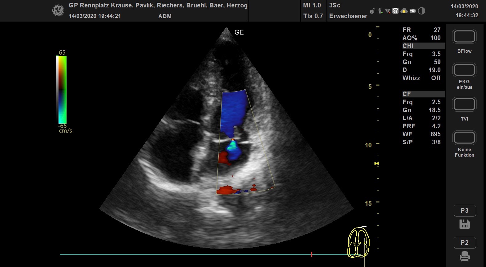
Echocardiography is the examination of the heart by ultrasound. Alongside the electrocardiogram (ECG), echocardiography is one of the most important technical non-invasive methods for examining the heart and is now an indispensable part of cardiological diagnostics. It is also a valuable diagnostic tool for general internists and rheumatologists.
A distinction is made between examining the heart through the chest wall (transthoracic echocardiography, TTE) and examining it from the oesophagus, for which a tube has to be swallowed, similar to a gastroscopy (transoesophageal echocardiography, TEE).
Echocardiography allows a detailed, non-invasive and gentle examination of the heart within a very short time. The size of the individual heart chambers, the pumping function and the function of the individual heart valves are clearly visible. Heart defects can also be detected quickly and reliably. Echocardiography can even detect heart defects in an unborn child. Certain heart defects or changes in the heart valves cannot be seen well enough with TTE, so a TEE is sometimes needed for a final diagnosis.
The examination through the chest is done from the parasternal position, i.e. next to the breastbone (sternum), and from the apical position, i.e. from the tip of the heart. Further positions are subcostal, where the ultrasound beam first passes through the liver and shows, for example, the width of the hepatic veins before they enter the inferior vena cava and the inferior vena cava before it enters the right atrium, and suprasternal. The short form “echo” usually refers to transthoracic echocardiography.
The different techniques allow the following to be examined: the 2D image shows a black-and-white cross-section in real time, so that heart function can be assessed live. In particular, the size of the heart chambers, the valve function and the pumping function of the heart are clearly visible, and it is possible, for example, to determine the ejection fraction (the proportion of blood pumped out of the heart chamber with each beat). The ascending aorta and the aortic arch can also be assessed in the parasternal long-axis view and in the suprasternal view, for example if an aortic dissection is suspected. PW and CW Doppler are mostly used from the apical position to measure the speed of blood flow, especially through the valves, in order to quantify valve stenoses, but also to determine pulmonary artery pressure or to assess the diastolic function of the left ventricle.
Colour Doppler helps to locate and quantify valve regurgitation and stenosis. In a different setting, colour Doppler is optimised for tissue and shows the movement of the heart wall. Ultrasound from the oesophagus is needed less often. For this, the patient has to swallow a tube with the ultrasound probe at its end, hence the nickname “swallow echo”. This method gives a better view of certain parts of the heart and of the thoracic aorta, and small blood clots, especially in the left atrial appendage, are detected more reliably.
A “stress echo” (stress echocardiography) is an echocardiography performed under stress. The most common reason for a stress echo is a suspected circulatory disorder due to coronary heart disease.
There are two ways of applying stress. In a mechanical stress echo, the patient lies on their left side on a bicycle ergometer and pedals against slowly increasing resistance while the doctor performs the ultrasound examination of the heart. In a pharmacological stress echo, the patient is given a medicine intravenously (usually dobutamine, or dipyridamole with atropine, or adenosine). Whatever the type of stress, the examination is carried out in several stages. First, images of the left ventricle are recorded at rest. The stress is then increased in several stages until a stopping criterion is met. Stopping criteria are reaching the target heart rate, the patient reporting chest pain, or a wall motion abnormality becoming visible on ultrasound. If the patient reports chest pain or a wall motion abnormality is seen, this strengthens the suspicion of coronary heart disease.